
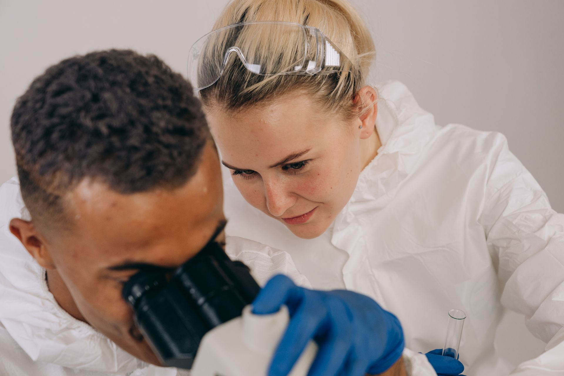
Histopathology
Haematoxylin & Eosin (H&E)
Haematoxylin and eosin is the routine tissue stain: nuclei are blue-purple and cytoplasm and connective tissue pink. It is the basis of almost every histopathology diagnosis, including tumour type, grade and margins.
Guideline: CAP Cancer Protocols / ICCR datasets; WHO Classification of Tumours (5th ed.)
Patient
Specimen & Request
Choose stain protocol
Histopathology
Haematoxylin & Eosin (H&E)
CAP Cancer Protocols / ICCR datasets; WHO Classification of Tumours (5th ed.)
Specimen
Microscopy
Prognostic Parameters
Finished reviewing the findings?
Smear Camera — teach STAINIC to read H&E
Snap the smear, film or microscope field with your phone camera, or upload a microscope camera image. Photos help STAINIC learn stain patterns so it can suggest findings in future.
Mobile data / Wi-Fi
Each photo (1-12 MB) is uploaded over your internet connection. On mobile data this uses your data plan and may incur charges; prefer Wi-Fi for many photos.
How AI will use photos
Photos are kept in a separate STAINIC image library (not your storage quota) to build future machine-learning models that recognise stain patterns and suggest findings. No AI analysis runs on them today and nothing is auto-filled yet.
Disclaimer
Photograph only the smear / film / field — never patient names, labels, faces or documents. Photos are not linked to any patient. Future AI suggestions will be decision-support only and must always be verified by a qualified pathologist; they are not a diagnosis or treatment advice.